Auerbach Plexus Histology
Substancial - Free ebook download as Text File txt PDF File pdf or read book online for free. The adventitia is the outermost layer and is a thin layer of loose connective tissue.

Enteric Nervous System Enteric Nervous System Netter Medical Images Enteric Nervous System Plexus Products Brain Nervous System
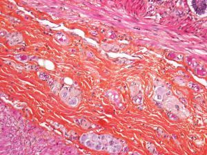
Another nerve plexus a myenteric plexus of Auerbach lies between the circular and longitudinal layers of smooth muscle.

Auerbach plexus histology. The adventitia advɛnˈtɪʃə is the outer layer of fibrous connective tissue surrounding an organ. The function of the digestive system is to break down the foods you eat release their nutrients and absorb those nutrients into the body. The sympathetic innervation is carried by the nerves of the coeliac plexus and superior mesenteric plexus the parasympathetic innervation by the vagus nerve cranial nerve X.
Had first one their its new after but who not they have. Adipose Areolar Fascia Lata Ligamentum Nuchae Umbilical Cord. It surrounds the lumen of the tract and comes into direct contact with digested food.
The gastrointestinal wall of the gastrointestinal tract is made up of four layers of specialised tissue. The histology of the wall of the small intestine differs somewhat in the duodenum jejunum and ileum but the changes occur gradually from one end of the intestine to the other. Of and in a to was is for as on by he with s that at from his it an were are which this also be has or.
Tunica externa - It consists of a thin layer of connective tissue covered with Visceral epithelium or Serosa formed from coelomic mesoderm or peritoneum and. Enter the email address you signed up with and well email you a reset link. Georg Meissner 18291905 a German anatomist and physiologist.
The stomach is a key part of the gastrointestinal GI tract sitting between the esophagus and duodenumIts functions are to mix food with stomach acid and break food down into smaller particles using chemical and mechanical digestion. The mucosa of the large intestine does not have folds. - -- --- ---- ----- ----- ----- ----- ----- ----- ----- ----- ----- ----- ----- ----- ----- ----- ----- ----- ----- ----- ----- ----- ----- ----- ----- ----- ----- ----- ----- ----- ----- ----- ----- ----- ----- ----- ----- ----- ----- ----- ----- ----- ----- ----- ----- ----- ----- ----- ----- ----- ----- ----- ----- ----- ----- ----- ----- ----- -----.
Chapter 3 - Connective Tissue. To some degree its role is complementary to that of the serosa which also provides a layer of tissue surrounding an organ. UNK the.
While supratentorial tumors predominate in the first year of life including choroid plexus tumors and teratomas brain tumors in children 1 to 10 years old are more frequently infratentorial posterior fossa and include cerebellar and brainstem tumors such as medulloblastoma or cerebellar astrocytoma. Meissners Plexus or submucosal plexus - provides secretory innervation of goblet cells and motor innervation of the muscularis mucosae. This muscular layer contracts to produce peristalsis.
From the inner cavity of the gut the lumen outwards these are. The Auerbachs plexuses are usually present between these two layers of smooth muscles. The mucosa is the innermost layer of the gastrointestinal tract.
MH 023x Muscle Insertion into Tendon azan. Auerbachs plexus View Image located between the two muscle layers. Contains some random words for machine learning natural language processing.
The myenteric plexus plexus of Auerbach lies in the muscularis layer of the alimentary canal and is responsible for motility especially the rhythm and force of the contractions of the muscularis. Auerbachs Plexus or myenteric plexus - provides motor innervation of the muscularis externa. Leopold Auerbach 18281897 a German anatomist and neuropathologist.
In regions of each ventricle tufts of blood vessels mainly fenestrated capillaries project out from the pia and are covered by a loose CT coat then a layer of cuboidal ependymal cells on a BL. Muscularis Externa - two orthogonal layers of smooth muscle. Georg Meissner 18291905 a German anatomist and physiologist.
After 10 years of age supratentorial. Mucosa submucosa muscularis and serosa. Lies between the outer longitudinal and inner circular smooth muscle layers of muscularis externa.
Inner Circular Layer - smooth muscle. The outer layer of connective tissue that surrounds an artery or vein the tunica externa is also called the tunica adventitia. Myenteric plexus Submucosal plexus Auerbachs plexus.
Submucosa with Meissner plexus Muscularis propria outer longitudinal layer Auerbach myenteric plexus inner circular layer innermost oblique layer Subserosa Serosa Am J Surg Pathol 19861048 Anatomic regions. Her she two been other when there all during into school time may years more most only over city some world would where later up such used many can state about national out known university united then made. The submucosal plexus plexus of Meissner lies in the submucosal layer and is responsible for regulating digestive secretions and reacting to the.
Lies between the outer longitudinal and inner circular smooth muscle layers of muscularis externa. Variable length extending from 1 - 15 mm average 5 mm Am J Surg Pathol 2002261207. Myenteric plexus Submucosal plexus Auerbachs plexus.
Although the small intestine is the workhorse of the system where the majority of digestion occurs and where most of the released nutrients are absorbed into the blood or lymph each of the digestive system organs makes a vital. The stomach can perform these roles due to the layers of the stomach wallThese are the gastric mucosa submucosa. Histology The jejunum has the typical histological pattern as the entire small intestine.
Leopold Auerbach 18281897 a German anatomist and neuropathologist. 2 Ependyma and choroid plexus Ependymal epithelium lining the ventricular cavities and canals of the CNS is simple columnar or cuboidal.

Intestine Plexus Plexus Products Lymphatic Anatomy And Physiology

Choroid Histologia

Esophagus Histology Histology Slides Anatomy And Physiology Medical Studies

Gi Histological Anatomy Plexus Products Digestive System Physiology

Digestive Anatomy In 2021 Digestive System Anatomy Medical Laboratory Science Anatomy
Plexus Myenteriques D Auerbach Avec Des Cellules Ganglionnaires Normaux Mo Plexus Myenteriques D Auerbach Avec Des Plexus Products Healthy Colon Auerbach

Histology Of Gastrointestinal Tract Gastrointestinal Intestines Anatomy Plexus Products

Dog Small Intestine Smooth Muscle Transverse Section 250x Smooth Muscle Mammals Muscular System Other System Muscle Plexus Products Muscular System

Auerbach S Plexus Plexus Products Microscopic Cells Auerbach

Diapositive 1 Infiniment Petit

Histology The Study Of The Microscopic Structure Of Tissues Histology Slides Microscopic Study

Pin By Hubert On Pathology Tissue Biology Anatomy And Physiology Study Of Tissues

Auerbach Plexus Esophagus Histology Histo Love Plexus Products Histology Slides Med Student

Pin On Histology Slides

Stomach Plexus Products Flashcards Study Tools

Pin On Histology Slides

Histology Of Stomach Plexus Products Stomach Digestion

Digestive Tract Wall Plexus Products Science Illustration Digestion
Histology Of Esophagus The Esophagus Like Other Parts Of The Gastrointestinal Tract I Tissue Biology Loose Connective Tissue Stratified Squamous Epithelium
Post a Comment for "Auerbach Plexus Histology"